


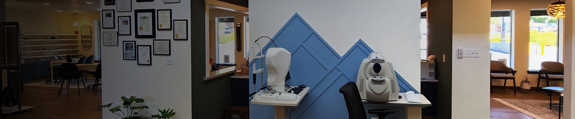
OUR TECHNOLOGY: RETINAL IMAGING
The eyes are the window to the soul and the retina is the window to your internal health. Looking at the arteries and veins in the retina can reveal diabetes, high blood pressure, high cholesterol, lupus, leukemia, sarcoidosis, cat scratch fever, medication use like tamoxifen and Plaquenil, and the list goes on. A dilated eye exam reveals the entire arena to the doctor in the moment, but a digital renal image lets you see inside your own eyes. It also provides a lasting record for future comparison, at a level of magnification beyond what a dilated exam can achieve.
Our retinal imaging system, the Eidon Ultra Widefield Scanning Laser Ophthalmoscope (SLO) is actually more than a camera. SLO imaging can show any changes in blood vessels from diabetes before they could be viewed on the surface. The autofluorescence feature can image macular degeneration and indicates the tissue health below the retina to help predict future vision changes and allow for earlier intervention. Retinal imaging is part of our Standard of Care and Drs. Ferris and Loeb recommend it as part of your examination. You never know what your photos will reveal about your health!
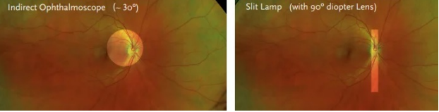
Images for comparison: Normal eye with undilated exam: Direct Ophthalmoscope (handheld device) or with Slit Lamp microscope (The bright area is the field of view seen with each device.)
Normal eye with our Eidon Ultra Widefield SLO image:

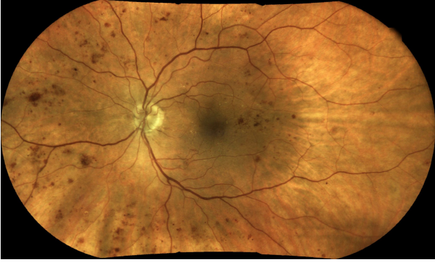
Eye with diabetic retinopathy:
Quick Links

All Eye
Care Services

Keep
In Touch
Contact Info
Hours of Operation
- Monday 8:30 AM - 5:00 PM
- Tuesday 8:30 AM - 5:00 PM
- Wednesday 10:00 AM - 6:00 PM
- Thursday 8:30 AM - 5:00 PM
- Friday 8:30 AM - 5:00 PM
- Saturday Closed
- Sunday Closed
© 2026 Rainier Eye. All rights Reserved. Accessibility Statement - Privacy Policy - Sitemap
Powered by: